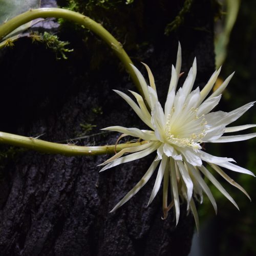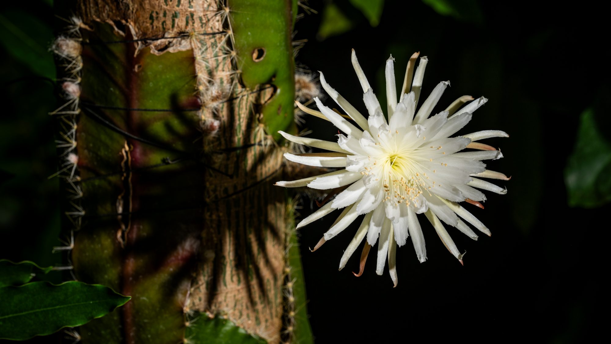
This winter we were excited to share the first ever multiple blooming of our tropical Amazonian moonflower – Strophocactus wittii. The three flowerings took place across nine days between 1-9 February and unlike Strophocactus wittii plants in the wild, which open at sunset and close by sunrise, ours all opened in the afternoon!
We set up a video livestream to track the blooms in anticipation of an evening flower – previous flowerings in 2021 and 2023 took place after closing hours, so we wanted to share what has become quite a celebrity plant with its many admirers and followers. However, much to the delight of our visitors, the flowers did not live up to their night-flowering moonflower name, and all chose to unfurl at various times in the afternoon: Flower A, began to unfurl on Thursday 1 February at approx 1.45pm; Flower B at approx. 12.45pm on Sunday 4 February and Flower C at 2.45pm on Friday 9 February.
Watch timelapse of moonflower C open
Enthusiastic visitors queued through the Glasshouses to catch sight of each flower unfurl as we shared the news on our social media and with local press. Some came to the Garden’s gates asking for updates, in the hope they could come and witness the flowering in person.


Although the flowers close just 12 hours after opening, these flowers had a much longer journey that was only just beginning. This multiple blooming meant more material was available for further study.
Moonflower goes under the microscope – scientists discover some of its inner secrets
With the plant producing three flowers rather than one single bloom, as in previous years, plant scientists at the University’s Sainsbury Laboratory (SLCU), located just a few yards from the Garden’s Glasshouses, were able to take the opportunity to dissect the flower and put fresh, live material under a powerful microscope.


The team were delighted as this tropical cactus is rare in cultivation and difficult to get hold of to study as its native habitat is the Brazilian rainforest. As far as we know, this is the first time the moonflower has been examined using an advanced low-temperature scanning electron microscope (cryo-SEM).
Dr Raymond Wightman, the Lab’s Microscopy Core Facility Manager explains that the cryo-SEM is a powerful imaging tool that enables live tissue to be imaged without damaging it: “In other forms of microscopy the samples have to be dried, heated or chemically processed before they can be imaged, which often destroys the native structure of the cells. Our cryo-SEM allowed us to image hydrated moonflower cells and capture the microscopic detail that provide us with insights into this unusual plant.”
“The plant is just a few yards from our microscope so it was able to be placed under a microscope within minutes of being collected.”
Kristina Buch, who is researching bumblebee visual ecology and petal epidermal (surface) cell wall architecture and development for her PhD, had booked the cryo-SEM for her own samples the morning after the first moonflower bloomed, but her plants were behind their predicted growth and not ready to image. So she was excited to discover the moonflower had bloomed the night before and was delighted with Wightman’s quick thinking to use the opportunity to examine the moonflower.
“Taking a closer look at the moonflower was a spontaneous wildcard that came about because of Ray’s suggestion.”
See the scientific process as the moonflower goes under the microscope
Film: courtesy of Sainsbury Laboratory, Cambridge University.
Kristina explains: “Over the past two years we have been using the cryo-SEM microscope to study petal surfaces across various plant species, but never those of a night-flowering plant, let alone of such a rare tropical specimen.
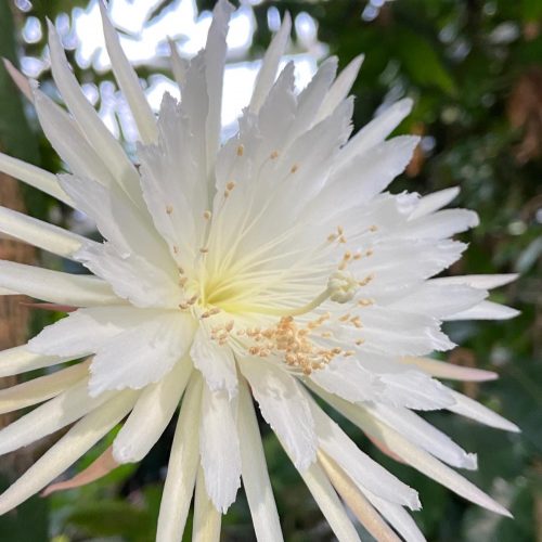
“This was an opportunity to take a closer look at the different petal-like structures layered into the flower, which we think are tepals – neither petals or sepals. We had some predictions, but we really had no idea what we might find. When examining the ultrastructure of the petal epidermal (surface) cell architecture, we found largely what we had hypothesised, with some surprises that deserve additional investigation.
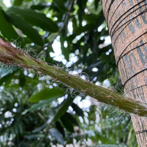
“We also want to have a closer look at the structural and biochemical parameters that might play a role in the UV-reflecting and absorbing properties of the petal and non-petal tissue. The outer layer of the flower’s receptacle (shoot) is covered by long fine fibres that we think might open some further routes of investigation, for example seeing if they create a more humid or protective micro-climate – either by shade or reducing evaporation or reflecting UV – or something else!”
Dr Wightman concludes that with the moonflower plant material being very scarce, they may have to wait for future flowers to pursue these additional questions: “We’ve taken hundreds of images that will take several weeks to analyse. There appears to be a few surprises that we’ll pay more attention to the next time it flowers….. whenever that may be.”
Discovering the dye held within the moonflower’s petals
CUBG’s Artist-in-Residence, Nabil Ali also took this rare opportunity to preserve the remaining petals of the moonflower. He used them to create a dye for his project (DYE – nature, myth and climate). The process involved heating a mixture of moonflower petals in vodka-water and grapefruit seed before adding a Potash mordant to create a light yellow tint.


While working with the flower, Nabil said that although the flower might look beautiful, it was one of ‘the most stubborn plants’ he has ever worked with! However, his hard work has paid off to produce lovely yellow colour, which he’s named ‘Moonflower yellow’.
If you would like to discover more about this enigmatic plant and previous flowerings, click on the links below.